


MTW European Type Trapezium Mill
Input size:30-50mm
Capacity: 3-50t/h
LM Vertical Roller Mill
Input size:38-65mm
Capacity: 13-70t/h
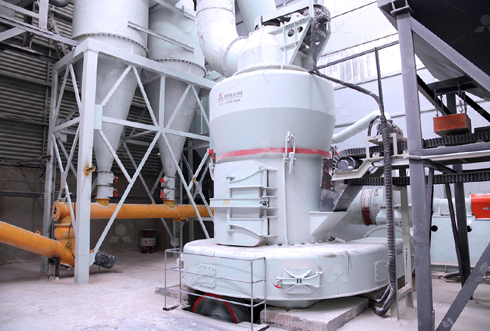
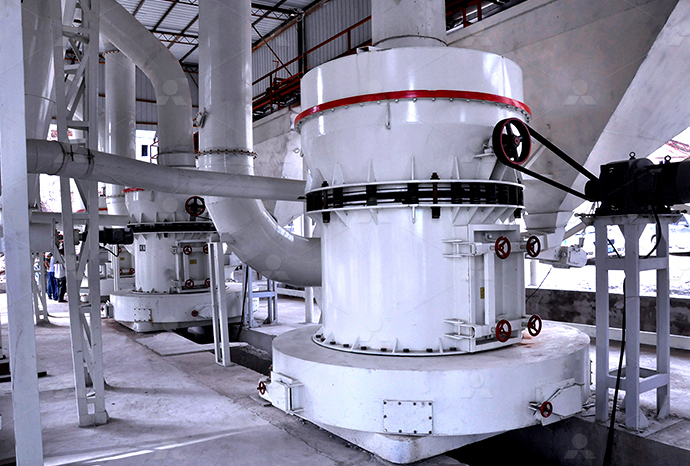
Raymond Mill
Input size:20-30mm
Capacity: 0.8-9.5t/h
Sand powder vertical mill
Input size:30-55mm
Capacity: 30-900t/h
LUM series superfine vertical roller grinding mill
Input size:10-20mm
Capacity: 5-18t/h
MW Micro Powder Mill
Input size:≤20mm
Capacity: 0.5-12t/h
LM Vertical Slag Mill
Input size:38-65mm
Capacity: 7-100t/h
LM Vertical Coal Mill
Input size:≤50mm
Capacity: 5-100t/h
TGM Trapezium Mill
Input size:25-40mm
Capacity: 3-36t/h
MB5X Pendulum Roller Grinding Mill
Input size:25-55mm
Capacity: 4-100t/h

Straight-Through Centrifugal Mill
Input size:30-40mm
Capacity: 15-45t/h
Xray chest film study of pneumoconiosis of wax stone miners and wax stone sculptors
.jpg)
Cardiothoracic ImagingImaging diagnosis of pneumoconiosis with
2023年5月1日 High resolution CT reflects pathological findings in pneumoconiosis and is useful for the diagnosis Pneumoconiosis such as silicosis, coal workers' pneumoconiosis, graphite 2022年11月3日 High resolution CT reflects pathological findings in pneumoconiosis and is useful for the diagnosis Pneumoconiosis such as silicosis, coal workers' pneumoconiosis, graphite Imaging diagnosis of pneumoconiosis with predominant nodular An improved CNNbased pneumoconiosis diagnosis method on Xray chest films is proposed to predict pneumoconiosis disease The CNN structure is decomposed from \ (5\times 5\) An Improved CNNBased Pneumoconiosis Diagnosis Method on X 2021年6月25日 A study (Zhu et al 2014) used normal (health) and abnormal (pneumociniosis) chest Xray films to extract texture features, and then used SVM algorithm to train and classify Pneumoconiosis identification in chest Xray films with CNNbased
AMFPnet: Adaptive multiscale feature pyramid network for
2024年8月1日 Automated pneumoconiosis classification in chest Xray images is challenging We propose an adaptive multiscale context module, to extract rich contextual features The method of radiographic diagnosis of pneumoconiosis has been internationally standardized; namely, each film is evaluated according to the profusion, shape, and size of small opacities Digital Xray images of pneumoconiosis and their evaluation2006年1月1日 In an individual who has a history of exposure to silica or coal dust, a finding of nodular or reticulonodular lesions at chest radiography or small nodules with a perilymphatic Pneumoconiosis: Comparison of Imaging and Pathologic FindingsIn this study, we proposed an ensemble learning approach to improve pneumoconiosis detection in chest Xrays (CXRs) using nine machine learning classifiers and multidimensional deep Detection and Visualisation of Pneumoconiosis Using an Ensemble
.jpg)
Imaging diagnosis of classical and new pneumoconiosis:
2021年3月10日 In this article, we demonstrate pathology and HRCT findings of asbestosis, uncommon pneumoconioses, and newly recognized pneumoconioses Compared to chest 2024年5月21日 Pneumoconiosis is an occupational lung disease caused by excessive exposure to respirable particles such as coal, silica and asbestos, and the disease caused by the inhalation of coal dust is also DLANet: dual lesion attention network for classification of 2010年9月15日 Request PDF Progression of Pneumoconiosis in Coal Miners after Cessation of Dust Exposure: A Longitudinal Study Based on Periodic Chest Xray Examinations in Hokkaido, Japan The progression Progression of Pneumoconiosis in Coal Miners after Cessation of In order to clarify the natural course of chest Xray findings of coal miners’ pneumoconiosis, we performed a longitudinal study based on Xray findings following the changes in subjects with pneumoconiosis that resulted from coal mining This is the first long term study on the natural course of pneumoconiosis in coal miners in Japan The imProgression of Pneumoconiosis in Coal Miners after Cessation of
.jpg)
Progression of pneumoconiosis in coal miners after cessation of
The progression of pneumoconiosis was observed after the cessation of dust exposure, especially during the first 10 years of retired coal miners in Hokkaido, Japan BACKGROUND The progression rate of pneumoconiosis in retired coal miners over ten years has not been studied in Japan METHODS A retrospective longitudinal study was undertaken using chest Xrays of Three randomized crossfold datasets of original database 32 Method Gao Huang et al developed DenseNet121, a CNN with dense connections between layers that was trained on the ImageNet database of 1000 classes, in 2017 []The four dense blocks were utilised to make these connections, which consisted of joining them in such a way that their output sizes were similarDetection and Visualisation of Pneumoconiosis Using an 2019年12月12日 The diagnosis of pneumoconiosis is very complex and cumbersome, which relies mostly on doctor’s medical knowledge and clinical reading experiences of Xray chest filmAn Improved CNNBased Pneumoconiosis Diagnosis Method on Xray Chest Film2021年6月25日 Pneumoconiosis is one of the most universal and severe occupational diseases in the world today The diagnosis of pneumoconiosis mainly depends on the doctors’ analysis of chest Xray films However, diagnostic accuracy is related to a doctor’s expertise Recent research has suggested that deep convolutional neural network (CNN) could better identify Pneumoconiosis identification in chest Xray films with CNN
.jpg)
Imaging diagnosis of pneumoconiosis with predominant nodular
2022年11月3日 The radiological patterns of known pneumoconiosis have been changing in recent years The basic pathology in pneumoconiosis is the presence of dust macules, mixed dust fibrosis, nodules, diffuse interstitial fibrosis, and progressive massive fibrosis These pathologic changes can coexist in dustexposed workers High resolution CT reflects pathological findings 2006年1月1日 Pneumoconiosis may be classified as either fibrotic or nonfibrotic, according to the presence or absence of fibrosis Silicosis, coal worker pneumoconiosis, asbestosis, berylliosis, and talcosis are examples of fibrotic pneumoconiosis Siderosis, stannosis, and baritosis are nonfibrotic forms of pneumoconiosis that result from inhalation of iron oxide, tin oxide, and Pneumoconiosis: Comparison of Imaging and Pathologic FindingsTHE early detection and correct classification of pneumoconiosis is still one of the difficult problems in diagnostic roentgenology It may, therefore, be of interest to summarize recent observations concerning the diagnosis and differential diagnosis of the disease, and to discuss its classification from the point of view of the clinical radiologist Pneumoconiosis has been The Xray Aspects of Pneumoconiosis Radiology RSNA Setting: Report of a workshop on Occupational Lung Diseases, IUATLD Respiratory Disease Section, Bangkok 1998 Objective: To describe the role of chest radiography in the diagnosis of pneumoconiosis in clinical practice Materials and methods: Pneumoconiosis, defined as the accumulation of dust in the lung and tissue reaction to its presence, is diagnosed and Chest radiography in the diagnosis of pneumoconiosis PubMed
.jpg)
Deep Learning Models of MultiScale Lesion Perception Attention
2024年6月5日 Accurate prediction of pneumoconiosis is essential for individualized early prevention and treatment However, the different manifestations and high heterogeneity among radiologists make it difficult to diagnose and stage pneumoconiosis accurately Here, based on DR images collected from two centers, a novel deep learning model, namely Multiscale American Roentgen Ray Society Images of Pneumoconiosis chest x ray All Images Xrays Echo Ultrasound CT Images MRI; Ongoing Trials at Clinical Trialsgov US National Guidelines Clearinghouse NICE Guidance FDA on Pneumoconiosis chest x ray CDC on Pneumoconiosis chest x ray Pneumoconiosis chest x ray in the news Blogs on Pneumoconiosis Pneumoconiosis chest x ray wikidoc2024年5月29日 Background Pneumoconiosis, a chronic disease stemming from prolonged inhalation of dust particles, stands as a significant global burden of occupational diseases This study aims to investigate the survival outcomes of pneumoconiosis patients in Huangshi city, China, while also evaluating the disease burden on afflicted patients Methods Data for this Survival and disease burden analyses of occupational pneumoconiosis 2021年2月1日 Request PDF Automated detection of pneumoconiosis with multilevel deep features learned from chest XRay radiographs Early detection of pneumoconiosis in XRays has been a challenging task Automated detection of pneumoconiosis with multilevel deep features
.jpg)
AMFPnet: Adaptive multiscale feature pyramid network for
2024年8月1日 For pneumoconiosis screening, chest Xray images (CXR) are commonly used due to low radiation dosage, wide availability and relatively low cost [4, 5]According to International Labour Organization (ILO) guidelines [6] based on the profusion level of small opacities observed in the lung, pneumoconiosis can be grouped into four main categories, 2009年5月1日 Digital chest imaging has replaced film chest radiographs in many centers, but the International Labour Organization classification system, which is the most widely used system for recognition and Comparison of Digital Radiographs with Film Radiographs for Background: The progression rate of pneumoconiosis in retired coal miners over ten years has not been studied in Japan Methods: A retrospective longitudinal study was undertaken using chest Xrays of 1091 pneumoconiosis subjects in Hokkaido, Japan between 1985 and 2005 Results: The final numbers of subjects were 207 (19% of the entry) after 1 decade and 85 Progression of pneumoconiosis in coal miners after cessation of The abnormality on a chest Xray of the lung is signified by an increase or decrease in density areas The chest Xray lung abnormalities with increased density are also known as pulmonary opacities Pulmonary opacities have three major patterns: ComputerAided Diagnosis of Coal Workers’ Pneumoconiosis in Chest
.jpg)
Epidemiology of coal miners’ pneumoconiosis and its social
2023年4月28日 Epidemiology of coal miners’ pneumoconiosis and its social determinants: An ecological study from 1949 to 2021 in China Background: Pneumoconiosis is the most widely distributed occupational disease worldwide China is currently the largest coal producer and consumer and the country with the most coal miners and cases of coal workers' 2013年4月2日 A wide spectrum of pulmonary complications occurs in patients with pneumoconiosis Those complications include chronic obstructive pulmonary disease, hemoptysis, pneumothorax, pleural disease, tuberculosis, Complications of pneumoconiosis: Radiologic overview2002年2月1日 The aim of this study was to develop a screening system of chest radiographs of miners with pneumoconiosis Chest radiographs were of coal mine or silica dust exposed miners participating in a A screening system for the assessment of opacity profusion in chest 2023年9月22日 Presently, the process of diagnosing occupational pneumoconiosis heavily relies on comparing patients’ chest Xray images to a standardized reference film However, this comparison method fails to optimally assist with early diagnoses, disease assessments, and prognosis guidancePrevention and Treatment of Pneumoconiosis in the Context of
.jpg)
Imaging diagnosis of classical and new pneumoconiosis:
In a study of the deposition and clearance of asbestos in rats using radioactive tracer techniques, autoradiographs of lung sections indicated that alveolar deposition was relatively uniform initially, that over a period of several months the uniform distribution changed to one in which fibers accumulated in foci that are mainly subpleural, and that these foci acted as centers for the Early detection of pneumoconiosis in XRays has been a challenging task that leads to high inter and intrareader variability Motivated by the success of deep learning in general and medical image classification, this paper proposes an approach to automatically detect pneumoconiosis using a deep f Automated detection of pneumoconiosis with multilevel deep features This review describes the prevalence of pneumoconiosis among coal miners, including underground, surface, and former coal miners; primary and secondary prevention for this disease; measures of disease severity, including mortality and lung transplants; the role of accurate radiograph classification by B Readers; and other respiratory conditions caused by coal mine Current Review of Pneumoconiosis Among US Coal Miners2006年1月1日 Pneumoconiosis may be classified as either fibrotic or nonfibrotic, according to the presence or absence of fibrosis Silicosis, coal worker pneumoconiosis, asbestosis, berylliosis, and talcosis are examples of fibrotic pneumoconiosis Siderosis, stannosis, and baritosis are nonfibrotic forms of pneumoconiosis that result from inhalation of iron oxide, tin oxide, and Pneumoconiosis: Comparison of Imaging and Pathologic Findings
Research Status of Pathogenesis of Pneumoconiosis and Dust
2021年11月3日 Pneumoconiosis has become one of the biggest threats to the occupational health and life safety of mining workers in China The number of pneumoconiosis cases has continued to rise in recent years The main task of occupational health development is to study the pathogenesis of pneumoconiosis and to develop mine dust prevention and control 2021年2月1日 Early detection of pneumoconiosis in XRays has been a challenging task that leads to high inter and intrareader variability Motivated by the success of deep learning in general and medical image classification, this paper proposes an approach to automatically detect pneumoconiosis using a deep feature based binary classifierAutomated detection of pneumoconiosis with multilevel deep 2021年5月27日 Pneumoconiosis is prevalent worldwide, and has maintained a relatively high incidence in recent years[4,5] It remains a severe global public health issue due to the lack of prevention of dust in the workplace, the failure Pneumoconiosis: current status and future prospectsAn improved CNNbased pneumoconiosis diagnosis method on Xray chest films is proposed to predict pneumoconia disease and achieves higher accuracy and gives a good result in the diagnosis Pneumoconiosis is one of the most serious occupational diseases in China, which seriously endangers the health of most workers in dust environments The diagnosis of An Improved CNNBased Pneumoconiosis Diagnosis Method on Xray Chest Film
.jpg)
The Pneumoconioses Radiology Key
2016年2月28日 COAL WORKER’S PNEUMOCONIOSIS Coal worker’s pneumoconiosis is a compensable occupational disease in the United States This disease is particularly common in underground miners In studies of coal 2023年7月25日 Pneumoconiosis is any lung disease caused by the inhalation of organic or nonorganic airborne dust and fibers Patients usually encounter these inhalants in the workplace environment, and therefore it is known as an occupational disease The most frequently encountered types of pneumoconiosis are asbestosis, silicosis, and coal miner’s lung These Pneumoconiosis StatPearls NCBI BookshelfThis work raises two transfer learning patterns from “frozen layers” and “finetuned layers" to solve problems of pneumoconiosis identification and proposes some methods of image denoising, lung segmentation, and data amplification for preprocessing to improve image qualities Pneumoconiosis is one of the most universal and severe occupational diseases in the world Pneumoconiosis identification in chest Xray films with CNN 2024年7月2日 Background Pneumoconiosis has a significant impact on the quality of patient survival due to its difficult staging diagnosis and poor prognosis This study aimed to develop a computeraided diagnostic system for the screening and staging of pneumoconiosis based on a multistage joint deep learning approach using Xray chest radiographs of pneumoconiosis Deep learning pneumoconiosis staging and diagnosis system
.jpg)
Pneumoconiosis: Symptoms, Causes, Risk Factors, Management
2024年1月10日 Pneumoconiosis is a lung disease caused by inhaling dust particles in the work environment These particles can include asbestos, coal dust, or silica Symptoms include difficulty breathing Black lung disease refers to the type of pneumoconiosis often diagnosed in coal miners Unfortunately, pneumoconiosis is not curableThe presence of opacities and plaques on the chest Xray is evidence there is scar tissue in the lungs, and pleural plaques are a common indicator of asbestosrelated disease 3 The chest Xray and CT scan provide evidence of fibrotic lesions, likely resulting from asbestos fibers in the lungs, and the MRI FDGPET scan was negative, which suggests the diagnosis is unrelated to lung Educational Case: Pneumoconiosis PMC2023年2月14日 Conclusions: The study presents pneumoconiosis data in a mixed and large pneumoconiosis in coal miners after cessation of dust exposure: a longitudinal study based on periodic chest Xray A Descriptive Study of a Turkish Pneumoconiosis CaseSeries2022年5月25日 Computeraided diagnostic (CAD) systems can assist radiologists in detecting coal workers’ pneumoconiosis (CWP) in their chest Xrays Early diagnosis of the CWP can significantly improve workers’ survival rate The development of the CAD systems will reduce risk in the workplace and improve the quality of chest screening for CWP diseases This ComputerAided Diagnosis of Coal Workers’ Pneumoconiosis in Chest