


MTW European Type Trapezium Mill
Input size:30-50mm
Capacity: 3-50t/h

LM Vertical Roller Mill
Input size:38-65mm
Capacity: 13-70t/h
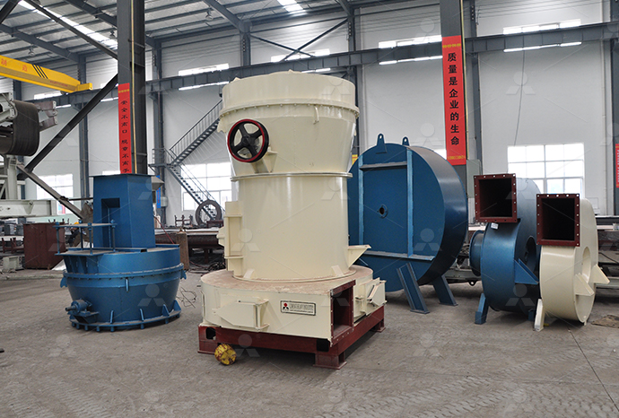
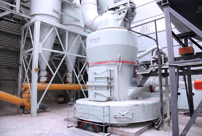
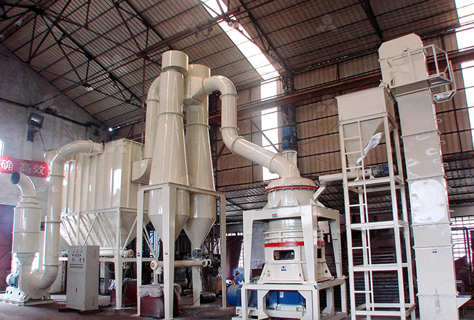
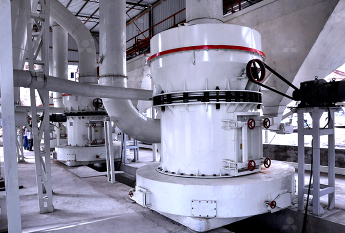
Raymond Mill
Input size:20-30mm
Capacity: 0.8-9.5t/h

Sand powder vertical mill
Input size:30-55mm
Capacity: 30-900t/h

LUM series superfine vertical roller grinding mill
Input size:10-20mm
Capacity: 5-18t/h
MW Micro Powder Mill
Input size:≤20mm
Capacity: 0.5-12t/h

LM Vertical Slag Mill
Input size:38-65mm
Capacity: 7-100t/h

LM Vertical Coal Mill
Input size:≤50mm
Capacity: 5-100t/h
TGM Trapezium Mill
Input size:25-40mm
Capacity: 3-36t/h

MB5X Pendulum Roller Grinding Mill
Input size:25-55mm
Capacity: 4-100t/h

Straight-Through Centrifugal Mill
Input size:30-40mm
Capacity: 15-45t/h
Limestone electron microscope
.jpg)
Scanning electron microscopy (SEMEDX) of limestone and burnt
In this paper, we present the importance of careful selection in the limestone and calcination process, which influences critical lime quality characteristicsThis study analysed the effect of three alkalizing soil amendments (limestone, Scanning electron microscop2012年12月1日 Morphological and optical observations of both the red and white limestones using petrographic microscopy, scanning electron microscopy (SEM), and highresolution Origin of the red colour in a red limestone from the Vispi Quarry 2022年9月19日 The microstructure changes in the limestone after acidification were studied via the wave velocity test and electron microscope scanning, and the damage deterioration mechanism was revealed The results show that the Microscopic Damage to Limestone under Acidic
Review on Electron Microscope Studies of Limestones
Electron microscope studies of limestones are reviewed based on own studies and on the evaluation of published data Further investigations should draw more attention to the 2021年11月12日 Polished thinsections prepared from the limestone samples were characterised by polarised light microscopy (PLM) using an Olympus BX53M optical The microstructural character of limestone and its influence on 2018年10月17日 A Cameca SX100 electron microprobe, operating at 15 kV voltage and 10 nA beam current with a 5 s counting time, coupled with EDS as well as a wave dispersive Elemental analysis of limestone by laserinduced breakdown A preliminary electron microscope study of fresh, unpolished fractured surfaces of 31 samples of limestone reveals features that correlate well with appearance under the optical microscope Surface morphology of some limestone types as revealed by
.jpg)
Review on Electron Microscope Studies of Limestones
Electron microscope studies of limestones are reviewed based on own studies and on the evaluation of published data R L Folk: Surface morphology of some limestone types as revealed by electron microscopy J Sediment Petrol 34, 144–155 (1964) Google ScholarDownload scientific diagram Scanning electron microscope (SEM) images of limestone (a), dolomite (b), chalcedonite (c) from publication: Successful Outcome of Phytostabilization in Cr(VI Scanning electron microscope (SEM) images of Download scientific diagram Scanning electron microscope (SEM) images of: a Basalt; b Limestone; and c Silica sand particles from publication: Geoelectrical Characterisation for CO2 Scanning electron microscope (SEM) images of: a An electron microscope is a microscope that uses a beam of electrons as a source of illumination They use electron optics that are analogous to the glass lenses of an optical light microscope to control the electron beam, for instance focusing them to produce magnified images or electron diffraction patterns As the wavelength of an electron can be up to 100,000 times smaller than Electron microscope Wikipedia
.jpg)
A quantitative scanning electron microscope study of evidence
A quantitative scanning electron microscope study of evidence for lichen weathering of limestone, Mendip Hills, Somerset Heather Viles, Many saxicolous lichens may cause physical and chemical weathering of limestone substrates, but their geomorphological importance has not been fully determinedDownload scientific diagram The examination of the limestone samples under polarized microscope shows Afine grained calcite (micrite), BDolomite and iron oxides, Cphosphate, Diron oxide and The examination of the limestone samples under polarized microscope 2013年5月23日 In this paper, we use scanning electron microscopy before and after salt weathering tests in cubic specimens of three limestone types (two grainstones and a travertine) in an attempt to built conceptual models that relate petrographic features and salt weathering susceptibility (represented by mass loss)Susceptibility of Limestone Petrographic Features to Salt For the electron microscope, an accelerating voltage of 100 kV produces a beam wavelength of close to 0004 nm The numerical aperture of the instrument is close to 0012, so take a minute and calculate the resolution of the electron microscope using the equation from earlier in this chapter (Hint: It’s 02 nmVisualizing Cells through Microscopy – Fundamentals of Cell

A quantitative scanning electron microscope study of evidence
Many saxicolous lichens may cause physical and chemical weathering of limestone substrates, but their geomorphological importance has not been fully determined A simple method of producing quantitative descriptions of lichen material and petrographic evidence of weathering using the SEM has been developed and tested on samples from the Mendip Hills, England 2018年6月9日 Electron microscope (EM) uses highenergy electron beam as probe instead of visible light The electrons have shorter wavelength and provides very highresolution capacity (01#160;nm) and 500,000 times magnification power It Electron Microscopy: Principle, Components, Optics and How the Great Pyramids of Giza were built has remained an enduring mystery In the mid1980s, Davidovits proposed that the pyramids were cast in situ using granular limestone aggregate and an alkali aluminosilicatebased binder Hard evidence for this idea, however, remained elusive Using primarily scanning and transmission electron microscopy, we compared a number of Microstructural Evidence of Reconstituted Limestone Blocks in the Electron microscope observations suggest that the hematite pigment has a diagenetic originThe red color is due to the and DeF are white limestone samples A, nanograins of calcite and hematite in red limestone B, selected Origin of the red colour in a red limestone from the
.jpg)
23: Instruments of Microscopy Biology LibreTexts
Whereas transmission electron microscopy requires very thin sections and allows one to see internal structures such as organelles and the interior of membranes, scanning electron microscopy can be used to view the surfaces of larger 2012年12月1日 Request PDF Origin of the red colour in a red limestone from the Vispi Quarry section (central Italy): A highresolution transmission electron microscopy analysis Hematite has been considered Origin of the red colour in a red limestone from the Vispi Quarry 2018年10月17日 Elemental analysis of limestone by laserinduced breakdown spectroscopy, scanning electron microscopy coupled with energy dispersive xray spectroscopy and electron probe microanalysis Muhammad Fahad 8,1, Zahid Farooq 2,3, Boynton R S 1980 Chemsitry and Technology of Lime and Limestone 2nd edn (New York: Wiley) p 592Elemental analysis of limestone by laserinduced breakdown 2023年8月19日 However, with an electron microscope, you can view it in 3D The exception is with stereo microscopes, which uses two eyepieces to create a 3D image Finally, a light microscope allows you to see the specimen exactly how it is, meaning in full color With an electron microscope, the image is seen in black and whiteLight Microscope vs Electron Microscope: 7 Main Differences

Scanning electron microscope images (backscattered electron
Scanning electron microscope images (backscattered electron mode; BSE) of polished oolitic limestone samples: ( a ) oolitic limestone 1 and ( b ) magnification of region bounded by dashed white 2021年11月12日 Polished thinsections prepared from the limestone samples were characterised by polarised light microscopy (PLM) using an Olympus BX53M optical microscope with an Olympus DP27 digital camera, and a Tescan MIRA II LMU scanning electron microscope with an energydispersive analytical system (Bruker AXS) (SEM–EDS)The microstructural character of limestone and its influence on Download scientific diagram Limestone powder (a) and cement (b) at SEM (scanning electron microscopy) from publication: Effect of limestone powder substitution on mechanical properties and Limestone powder (a) and cement (b) at SEM (scanning electron SEM stands for Scanning Electron Microscope An SEM is a type of electron microscope that uses a fine beam of focused electrons to scan a sample’s surface SEM images give insight into a sample’s surface topography; therefore, it creates 3D Different types of Microscopes – light microscope, electron
.jpg)
Electron Microscopy: Principle, Components, Optics and
2023年1月1日 An electron microscope (EM) uses a high energy electron beam aa s probe instead of visible light The electrons have a shorter wavelength and provide a very highresolution capacity (01#160;nm) and 500,000 times Observing chalk under a microscope is another fun experiment as you begin in microscopy! Read on some researchers, the majority of prehistoric cave paintings (between 40,000 and 10,000 BC) were made using chalk and Chalk under a Microscope Procedures and Observations2022年9月19日 In order to study acid damage’s effect on this kind of rock, limestone samples were acidified for 0 days, 5 days, 10 days, 15 days, and 20 days The microstructure changes in the limestone after acidification were Microscopic Damage to Limestone under Acidic 2018年8月23日 The history of the electron microscope dates back to early twentieth century when the first electromagnetic lens was developed This opened the door of possibility to use the principles of the History of the Electron Microscope NewsMedical
.jpg)
A Universal Classification Scheme For the Microcrystals That
2015年10月1日 Presented here is a global assessment of limestone microporosity that is based on scanning electron microscope, crystal size distributions, helium porosimetry, and mercuryinjection capillary 2022年5月19日 Magnetic fields were used as lenses for the electrons With these discoveries, the first electron microscope was later developed by Ernst Ruska and Max Knolls in 1931 and modified into a Transmission Electron Microscope (TEM) by Ernst Ruska along with the Sieman’s company, in 1933Transmission Electron Microscope (TEM) Definition, Principle, 2022年9月20日 Coral reef limestone (CRL) is a biomass limestone formed by reefbuilding corals and other biological skeletons Electron microscope scanning of CGL: (a) magnification: 227kx; (b) magnification: 51× The surface of the CGL specimens contained a large number of macroscopic pores (Fig 1 c)Mesoscopic damage evolution of coral reef limestone based on A preliminary electron microscope study of fresh, unpolished fractured surfaces of 31 samples of limestone reveals features that correlate well with appearance under the optical microscope Smooth, granual, acicular, and rhombohedral surfaces are recognizable; some are Surface morphology of some limestone types as revealed by electron

Susceptibility of Limestone Petrographic Features to
2013年5月23日 Request PDF Susceptibility of Limestone Petrographic Features to Salt Weathering: A Scanning Electron Microscopy Study Salt weathering is a major erosive process affecting porous materials in 2019年11月22日 Reflected Light Microscopy as an Efficient and CostEffective Method for the Detection of Calcareous MicroFossils, an Example from the Much Wenlock Limestone Formation, Shropshire, UK(PDF) Reflected Light Microscopy as an Efficient and Cost 2023年4月13日 In conclusion, electron microscopy instrumentation is intricate and depends on a variety of cuttingedge parts and computer systems Highresolution imaging of materials and biological samples at the atomic and Electron Microscopy: Types, Instrumentation, The Scanning Electron Microscope images of the LCC blend can be found in Fig 3 Fine silica sand with particle size of 120180 μm was used to control the matrix fracture toughness ( Wu et al Scanning Electron Microscope images of limestone

Investigations of a Cretaceous limestone with spectral induced
2016年11月17日 Studies that combine scanning electron microscopy and the IP method show that the intensity of the IP effect may vary with the volumetric content of disseminated conductive minerals in the rocks An account of the early history of scanning electron microscopy has been presented by McMullan [2] [3] Although Max Knoll produced a photo with a 50 mm objectfieldwidth showing channeling contrast by the use of an electron beam scanner, [4] it was Manfred von Ardenne who in 1937 invented [5] a microscope with high resolution by scanning a very small raster with a Scanning electron microscope WikipediaThe technique is here applied to an oolitic limestone and a carbonate laminite to illustrate its application as a tool to study carbonate porosity and diagenesis Highresolution large area scanning electron microscopy: an imaging tool for porosity and diagenesis of carbonate rock systems / Buckman, Jim; Charalampidou, ElliMaria Highresolution large area scanning electron microscopy: an the transmission electron microscope (TEM) is used to examine thin slices or sections of cells or tissues close tissue A group of similar cells that carry out the same function, eg muscle tissueElectron microscopes Cell structure Edexcel BBC

Better, Faster, Cheaper: Recent Advances in Cryo–Electron Microscopy
Cryo–electron microscopy (cryoEM) continues its remarkable growth as a method for visualizing biological objects, which has been driven by advances across the entire pipeline Developments in both singleparticle analysis and in situ tomography have enabled more structures to be imaged and determined to better resolutions, at faster speeds, and with more scientists having 2024年4月3日 Scanning electron microscopes (SEMs) Most of the funky electron microscope images you see in books—things like wasps holding microchips in their mouths—are not made by TEMs but by scanning electron microscopes (SEMs), which are designed to make images of the surfaces of tiny objects Just as in a TEM, the top of a SEM is a powerful electron gun that How do electron microscopes work? Explain that Stuff2024年1月2日 Electron microscopy has harnessed the power of physics to allow us to see beyond the limits imposed by visible light In this article, we explore how electron microscopy works, some of the common techniques and applications and Electron Microscopy Techniques, Strengths, Limitations and Applications2018年10月17日 Elemental analysis of limestone by laserinduced breakdown spectroscopy, scanning electron microscopy coupled with energy dispersive xray spectroscopy and electron probe microanalysisElemental analysis of limestone by laserinduced ResearchGate